ใครเข้าใจร pathway ด้านล่างช่วยอธิบายแบบคร่าวๆ หน่อยครับ ไม่ต้องละเอียดก็ได้ครับ ช่วยหน่อยครับผม ขอบคุณมากๆ ครับ
 คำอธิบายรูป
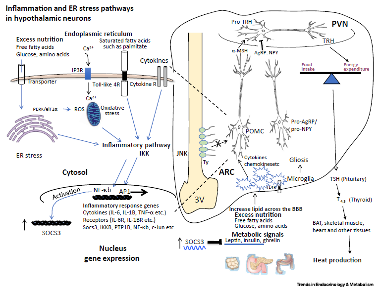
Figure 2. Hypothalamic Inflammation and Endoplasmic Reticulum (ER) Stress in Obesity. This figure depicts the hypothalamic inflammation observed in obesity that is temporally different from peripheral inflammation. Whereas peripheral inflammation is thought to develop secondary to diet-induced adiposity, rapid changes in hypothalamic inflammatory signaling cascades could be observed within 1 day of high-fat diet (HFD) consumption. For example, rodents display elevated markers of inflammation [40] and insulin resistance in the hypothalamic neuronal circuitry [71] within 24 h of HFD exposure, much earlier than the accumulation of adiposity. The central inflammation observed upon HFD consumption, at least in part, depends on the direct effect of saturated fatty acids on the hypothalamus rather than on the total calories consumed, and central infusion of saturated fatty acids to lean rodents mimics the hypothalamic insulin resistance and IKKb activation observed in animals exposed to HFD [72]. Hypothalamic inflammation may be stimulated by acute administration of saturated fatty acids, without induction of systemic inflammation. In parallel with their central effects in vivo, saturated fatty acids induce ER stress in neuronal cultures [25,73]. However, their proinflammatory effect is attenuated in cultured hypothalamic neurons, suggesting a role for non-neuronal cells in fatty acid-induced central inflammation [73] (Box 2). Consumption of a highcalorie diet results in activation of microglia in the ARC [74], and diet-induced hypothalamic gliosis (activation of the glial population) is also reported in humans [40]. The resident astrocytes and microglia are the innate immune cells of the CNS, and there is a dynamic interaction between them, including during central inflammation. Activated microglia can induce a subpopulation of astrocytes by secreting inflammatory mediators, including IL-1a, TNF, and C1q, which in turn blunt for example the neuroprotective capacity of the astrocytes, leading to neuronal death [75]. In contrast to the periphery, the central innate immune response in general cannot initiate adaptive immunity, consistent with the anti-inflammatory environment of the brain parenchyma as well as the physical limitations imposed by the BBB that block migration/communication of innate and adaptive immune cell components. Genetic and pharmacological suppression of neuroinflammation leads to metabolic improvements. These include potent weight loss and/or protection from diet-induced obesity by central or peripheral administration of a JNK2/3-specific inhibitor [76], targeted activation of the anti-inflammatory glucocorticoid receptor in GLP-1-positive cells [77], and neuron-, glia-, or hypothalamus-specific deletion of IKKb [21,78]. The ER establishes physical contacts with other organelles at membrane contact sites (not shown in the figure). Mitofusins (MFNs), for example, tether ER and mitochondria, and participate in central metabolic regulation (discussed in the main text). The figure also depicts the hypothalamus–pituitary–thyroid (HPT) axis as an example of a central pathway regulating energy balance. The HPT axis is a strong determinant of energy expenditure through the action of thyroid hormones in tissues such as skeletal muscle or BAT: a-MSH activates whereas AgRP and NPY block TRH expression in the PVN. TRH stimulates the pituitary gland to secrete TSH, which in turn leads to triiodothyronine (T3) and thyroxine (T4) production by the thyroid gland. Abbreviations: ARC, arcuate nucleus; BAT, brown adipose tissue; BBB, blood–brain barrier; PVN, paraventricular nucleus; TRH, thyrotropin releasing hormone; TSH, thyrotropin stimulating hormone; Ty, tanycytes; 3V, third ventricle.
ข้อมูลจาก งานวิจัย Cakir, I., Nillni, E.A.. 2019. Endoplasmic Reticulum Stress, the Hypothalamus, and Energy Balance. Trends in Endocrinology & Metabolism. XX. 1-14.
ลิงค์: DOI:
https://doi.org/10.1016/j.tem.2019.01.002
ขอบคุณมากครับ


ใครดูรูปนี้แล้วเข้าใจบ้างครับ (มีคำอธิบาย; กระบวนการการอักเสบและความเครียดของ ER ในเซลล์ประสาท hypothalamic)
คำอธิบายรูป
Figure 2. Hypothalamic Inflammation and Endoplasmic Reticulum (ER) Stress in Obesity. This figure depicts the hypothalamic inflammation observed in obesity that is temporally different from peripheral inflammation. Whereas peripheral inflammation is thought to develop secondary to diet-induced adiposity, rapid changes in hypothalamic inflammatory signaling cascades could be observed within 1 day of high-fat diet (HFD) consumption. For example, rodents display elevated markers of inflammation [40] and insulin resistance in the hypothalamic neuronal circuitry [71] within 24 h of HFD exposure, much earlier than the accumulation of adiposity. The central inflammation observed upon HFD consumption, at least in part, depends on the direct effect of saturated fatty acids on the hypothalamus rather than on the total calories consumed, and central infusion of saturated fatty acids to lean rodents mimics the hypothalamic insulin resistance and IKKb activation observed in animals exposed to HFD [72]. Hypothalamic inflammation may be stimulated by acute administration of saturated fatty acids, without induction of systemic inflammation. In parallel with their central effects in vivo, saturated fatty acids induce ER stress in neuronal cultures [25,73]. However, their proinflammatory effect is attenuated in cultured hypothalamic neurons, suggesting a role for non-neuronal cells in fatty acid-induced central inflammation [73] (Box 2). Consumption of a highcalorie diet results in activation of microglia in the ARC [74], and diet-induced hypothalamic gliosis (activation of the glial population) is also reported in humans [40]. The resident astrocytes and microglia are the innate immune cells of the CNS, and there is a dynamic interaction between them, including during central inflammation. Activated microglia can induce a subpopulation of astrocytes by secreting inflammatory mediators, including IL-1a, TNF, and C1q, which in turn blunt for example the neuroprotective capacity of the astrocytes, leading to neuronal death [75]. In contrast to the periphery, the central innate immune response in general cannot initiate adaptive immunity, consistent with the anti-inflammatory environment of the brain parenchyma as well as the physical limitations imposed by the BBB that block migration/communication of innate and adaptive immune cell components. Genetic and pharmacological suppression of neuroinflammation leads to metabolic improvements. These include potent weight loss and/or protection from diet-induced obesity by central or peripheral administration of a JNK2/3-specific inhibitor [76], targeted activation of the anti-inflammatory glucocorticoid receptor in GLP-1-positive cells [77], and neuron-, glia-, or hypothalamus-specific deletion of IKKb [21,78]. The ER establishes physical contacts with other organelles at membrane contact sites (not shown in the figure). Mitofusins (MFNs), for example, tether ER and mitochondria, and participate in central metabolic regulation (discussed in the main text). The figure also depicts the hypothalamus–pituitary–thyroid (HPT) axis as an example of a central pathway regulating energy balance. The HPT axis is a strong determinant of energy expenditure through the action of thyroid hormones in tissues such as skeletal muscle or BAT: a-MSH activates whereas AgRP and NPY block TRH expression in the PVN. TRH stimulates the pituitary gland to secrete TSH, which in turn leads to triiodothyronine (T3) and thyroxine (T4) production by the thyroid gland. Abbreviations: ARC, arcuate nucleus; BAT, brown adipose tissue; BBB, blood–brain barrier; PVN, paraventricular nucleus; TRH, thyrotropin releasing hormone; TSH, thyrotropin stimulating hormone; Ty, tanycytes; 3V, third ventricle.
ข้อมูลจาก งานวิจัย Cakir, I., Nillni, E.A.. 2019. Endoplasmic Reticulum Stress, the Hypothalamus, and Energy Balance. Trends in Endocrinology & Metabolism. XX. 1-14.
ลิงค์: DOI: https://doi.org/10.1016/j.tem.2019.01.002
ขอบคุณมากครับ